Content
BY CAROLINE HALL / 26 OCT 2020
https://aestheticsjournal.com/feature/case-study-delayed-onset-nodules

Here, we provide a case study of a dermal filler problem that was managed six months after the initial procedure.
Nodules with a slow onset
According to how soon after the first injection they occurred, complications brought on by filler injections are often categorised. A complication that develops days after therapy is categorised as an early event, whereas a reaction that develops weeks to years after injection is categorised as a delayed event.
Delayed onset nodules (DONs) are described in the literature as a palpable or visible unintended mass that develops close to or at the injection site of dermal filler. The variability between aetiology and clinical presentations can make delayed onset nodules (DONs) more difficult to address or prevent2. After being recognised by the practitioner, a DON can then be further classified as inflammatory or non-inflammatory. It may manifest weeks, or even years, after the initial treatment. The symptoms of an inflammatory nodule may include pain, tenderness, swelling, heat, and erythema4.
DONs are still a rare issue, but they are increasingly more frequently recognised. A more recent study from 2019 indicated that of 1,250 patients receiving filler treatment, the incidence of DONs was 1.0%. This contrasts with a four-year retrospective study of 4,320 filler treatments that was published in 20065, which found only a 0.6-0.8% rate of hypersensitive reactions.
A patient with an active autoimmune disease should be treated with extra caution because there isn’t enough solid information to say which patients are more likely to get DONs, but immuno reactive patients seem to experience them more frequently. There is mounting evidence linking cold and flu viruses to the development of delayed hypersensitivity reactions, with some studies suggesting an increase in reported reactions during the fall and winter “flu season.” 8, 9
In order to effectively handle the problem should it develop in their practise, practitioners must be aware of the potential for DONs to occur. It’s crucial to be able to explain and educate them about any potential side effects before beginning treatment.
Research case
Initial procedure
The patient was a 50-year-old woman who had never had any non-surgical treatments before. She visited because she was worried about jowl sagging and midface volume reduction. The patient was healthy and solely used medication for hormone replacement therapy. In order to support the mid-face, add volume there, and start to reduce the “sag” of the jowl area, the plan was to inject 1ml of a 20mg/ml hyaluronic acid, high G-prime, high cohesivity filler into each cheek after her initial appointment.
The patient’s skin was washed with Clinisept+ the day of the procedure, and a sterile wound care treatment pack was used. Lidocaine was present in the hyaluronic acid filler, and no topical anaesthetic was used to the skin. The MD Codes approach was used for injecting the hyaluronic acid filler. A 27 gauge needle was used to administer three 0.1 ml boluses to the periosteum at CK1, a 0.3 ml bolus to the periosteum at CK3, and a fanning method to provide 0.5 ml via a 25 gauge cannula to the deep dermis at CK3. The patient was satisfied with the treatment’s outcomes, and there were no immediate problems. Standard post-operative instructions were provided to the patient, including not to touch or massage the treated region, to stay out of severe heat or cold, to avoid strenuous activity, to refrain from wearing makeup for 24 hours, and to abstain from alcohol for 48 hours. The patient stated that she adhered to the aftercare instructions.
The patient came back eight weeks later and asked for more care. She requested an additional 0.5ml of hyaluronic acid filler to each side because her prior filler had settled well and she appreciated the extra volume to the mid-face area. Once more, the MD Codes approach was employed. A 27 gauge needle was utilised to inject 0.3 ml of the same hyaluronic acid filler into the periosteum at CK3 and 0.2 ml at CK2. The patient once more saw positive outcomes.
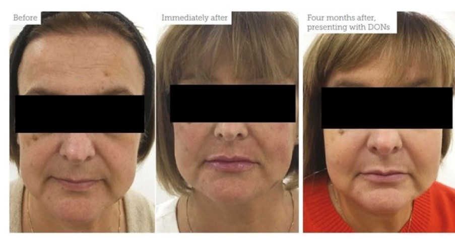
Figure 1 shows the patient’s condition prior to and right after the procedure, as well as four months later, when she presented with DONs. The DONs were not visible, but they were felt.
The difficulty
A call was received from the patient four months after the initial treatment to ask her to come in for a review since she had acquired swellings in the mid-face region.
During this session, it’s crucial to take a comprehensive approach to the patient by performing a physical exam and getting all the details about their medical history. Ask them things like if they had ever been sick before the incident, if their medication had changed, and if they had ever had any type of surgery. It would also be a good idea to find out whether the patient received any additional aesthetic treatments that you might not be aware of or if she had seen another doctor after her original appointment with you. Although the patient mentioned a period of increasing stress in her personal life, which was unlikely to be relevant in this case, she did not report any changes to their medical history or any periods of illness prior to the nodules developing.
Look for any skin issues or a proper barrier function when inspecting the face. The patient had a 2cm smooth lump on his right cheek and a 4cm smooth mass on his left cheek. Both cheeks were painful to the touch, but there was no erythema or heat to be felt. Although the nodules were not apparent to the naked eye, the patient said she felt embarrassed and worried that others might be able to see them.
The treatment strategy will depend on whether the nodules are inflamed or not, thus it is critical to ascertain this right away. A non-inflammatory nodule typically manifests as a painless, hard, cold, regularly formed lump without any redness. Pain, heat, erythema, and swelling are all symptoms of an inflammatory nodule.4,10 In this case, it was concluded that the nodules were inflammatory due to their delayed development and the patient’s tenderness and discomfort. There were no other skin issues found.
The patient was eager to try to correct the nodules without dissolving the filler because she had been so pleased with her results up until that point. Therapy options, including this possibility, were thoroughly addressed. We decided to try to diminish them using a variety of treatments, but since the possibility of having to dissolve them in the future couldn’t be ruled out, it was also decided to check in on progress periodically. The recommended course of treatment if the nodules had been non-inflammatory would have been to monitor and wait if they were not bothersome to the patient, or to dissolve with hyaluronidase if they were.
At first, the patient was instructed to take 500 mg of Clarithromycin twice daily for two weeks. The antibiotics were provided in accordance with the recommendations of the Aesthetic Complications Expert (ACE) Group3 and a 2020 study on delayed onset inflammatory nodules2. A 2009 study also supported the use of antibiotics as a first-line treatment for DONs. In a study of 55 patients who experienced reactions to polyacrylamide gels that were thought to be brought on by low virulence bacteria, those who initially received steroids or non-steroidal anti-inflammatory drugs (NSAIDs) as opposed to antibiotics had significantly worse prognoses than those in the antibiotics-first group.11
The patient was followed up frequently and had a face-to-face review two weeks after she finished the prescribed antibiotics. The nodules were 2 cm on the left and 1 cm on the right, having significantly shrunk. Although there was no erythema or heat, there was still some pain. Prednisolone 40mg once day for seven days followed by a reduction of 5mg daily for an additional seven days helped shrink the nodules. Although several studies support their use in effectively resolving inflammatory nodules,2,9 the use of steroids to reduce inflammatory conditions appears to be more at the prescribing practitioner’s discretion and is not documented in the ACE guideline algorithm for treatment of DONs in aesthetic medicine.
After another 14 days, The patient was reevaluated, and the nodules had disappeared after two weeks of antibiotics and two weeks of steroids. For the following four weeks, she was followed up with once a week, and throughout that time, reported no new problems.
Summary
Here is an illustration of effective DON management without the usage of hyaluronidase. It is well acknowledged that infection is the underlying cause of delayed inflammatory nodules, and it is recommended practise to employ antibiotics as the first line of treatment. Treatment with steroids, non-steroidal anti-inflammatories, and hyaluronidase are all supported in the literature as effective treatment choices. Practitioner variances will occur in subsequent treatments, and it is best practised on a case-by-case basis.
References
1. A review of adverse responses to dermal fillers by Lowe NJ, Maxwell CA, and Patnaik R. 2005;31(11 Pt 2):1616-1625 in Dermatology Surgery.
2. De Boulle K, Philipp-Dormston WG, Goodman GJ, et al. Worldwide Strategies for the Prevention and Treatment of Delayed-onset Adverse Events to Fillers Based on Hyaluronic Acid. Glob Open Plast Reconstr Surg. 2020;8(4):e2730.
3. S Bassett, M King, E Davies, and S King. Delayed Onset Nodule Management. Expert Panel on Aesthetic Complications. Date of review: 2017.
4. Management of problems with injectable soft tissue fillers, Sclafani A, Fagien S. Skin Surgery 2009;35:1672-1680
5. Dermal fillers in aesthetics: a review of adverse events and therapeutic options, Funt D, Pavicic T. Dermatol Clin Cosmet Invest. 2013;6:295-316.
6. Sadeghpour, N. A. Quatrano, L. M. Bonati, et al. Comparative incidence and risk assessment of delayed-onset nodules due to differently crosslinked hyaluronic acids. 2019;45:1085–1094 for Dermatologic Surgery.
7. Yang S, Ledon JA, Savas JA, et al. Review of pathophysiology, causes, and treatments for inflammatory nodules that develop after soft tissue usage in inflammatory nodules. AJCD. Am J Clin Dermatol. 2013;14(5):401–411.
8. Clinical experience with 11,460ml of smooth, extremely cohesive, viscous hyaluronic acid filler containing 20 mg/ml, by Humphrey, Carruthers, and Carruthers. Dermatol Surgery, September 2015; 41(9):1060-7.
9. Delayed hypersensitivity reaction to hyaluronic acid dermal filler during influenza-like sickness. Turkmani MG, De Boulle K, Philipp-Dormston WG. Dermatol Clin Cosmet Invest. 2019;12:277–283.
10. Late-Onset Inflammatory Reaction to Hyaluronic Acid Dermal Fillers, Bhojani-Lynch T. Glob Open Plast Reconstr Surg. 2017;5(12):e1532.
11. Host Tissue Interaction, Fate, and Hazards of Biodegradable and Nonbiodegradable Gel Fillers, Christensen LH. Skin Surgery 2009;35:1612–1619.